General description
This is our standard MRI protocol to visualize the eye and internal structures.
It contains isotropic 3D sequences, which can be use for an accurate assessment of the lesion’s dimensions and extent.
These scans are complemented with 2D sequences with a higher in plane resolution that allow for a more detailed radiological evaluation.
The scans are generally planned such that all relevant structures, e.g. tumour and adjacent optic nerve, are visible in a single slice.
In addition to these anatomical sequences, the protocol contains functional sequences, which provide biomarkers of the lesion’s vascularity (perfusion weighted imaging) and cellularity (diffusion weighted imaging). These scans are particularly valuable for the differential diagnosis, but can also be used as an early marker of treatment response.
References
- The technical rationale behind this protocol is described in
MRI of Uveal Melanoma

- The images acquired with this protocol have been further described/validated in the following studies:
- MR imaging characteristics of uveal melanoma with histopathological validation

- Comparison of Magnetic Resonance Imaging–Based and Conventional Measurements for Proton Beam Therapy of Uveal Melanoma

- MR-based follow-up after brachytherapy and proton beam therapy in uveal melanoma

- Eye-specific quantitative dynamic contrast-enhanced MRI analysis for patients with intraocular masses

- This protocol is furthermore the basis for our eye lid protocol and the protocol we use for the planning of ocular Proton Therapy.
Requirements
Hardware:
- 3 Tesla MRI
- Surface (receive) coil (we use a 47mm Philips receive coil)
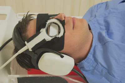
- A support for the head (we use the MaxSupportTM wide shaped (red) variant from Medeo)
- Preferably a mask/goggles for positioning of the coil (ours are made from radiotherapy thermoplastic material and Velcro)
- A power injector to administer a bolus of 0.1 mmol/kg gadoterate meglumine (Dotarem) 6s after the start of the dynamic scan, followed by a 20 ml injection of isotonic saline with an injection rate of 2 ml/s
- The affected eye is closed with a piece of tape and subsequently covered with a wet gauze to limit susceptibility artefacts at the air-cornea interface
Sequences:
- For the 3D sequences, we use a variable refocussing angle to allow for longer echo-trains without blurring, which substantially reduces the scan time. The Philips implementation is called BrainVIEW, but other vendors have similar options (eg. SPACE on Siemens and CUBE on GE).
- To increase the temporal and spatial resolution of the dynamic contrast enhancement (DCE) scan, the outer part of k-space is acquired over multiple dynamics. On Philips this is called ViewSharing, on Siemens TWIST and on GE TRICKS.
- For the quantification of the perfusion data (and T1 of the tumour), we determine the B1 with the DREAM sequence.
Protocol description
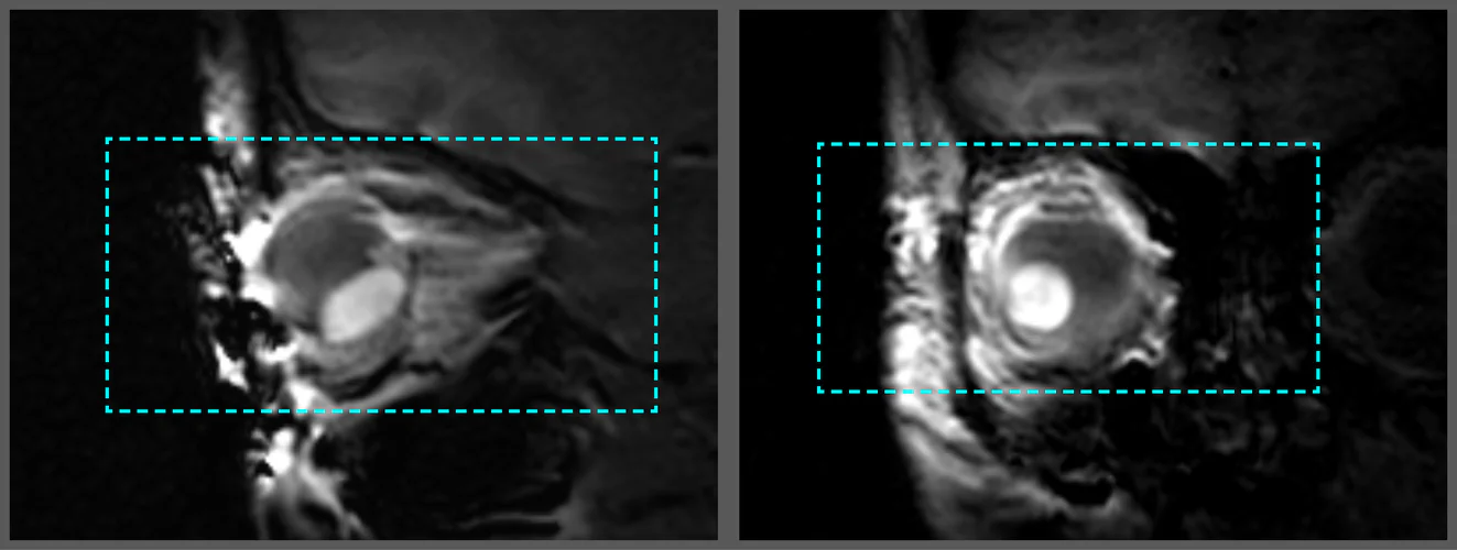
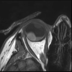
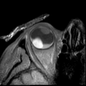
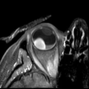
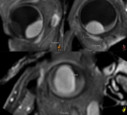
3DT1 SPIR Gd
The isotropic resolution allows for accurate 3D assessment of the lesion dimensions.
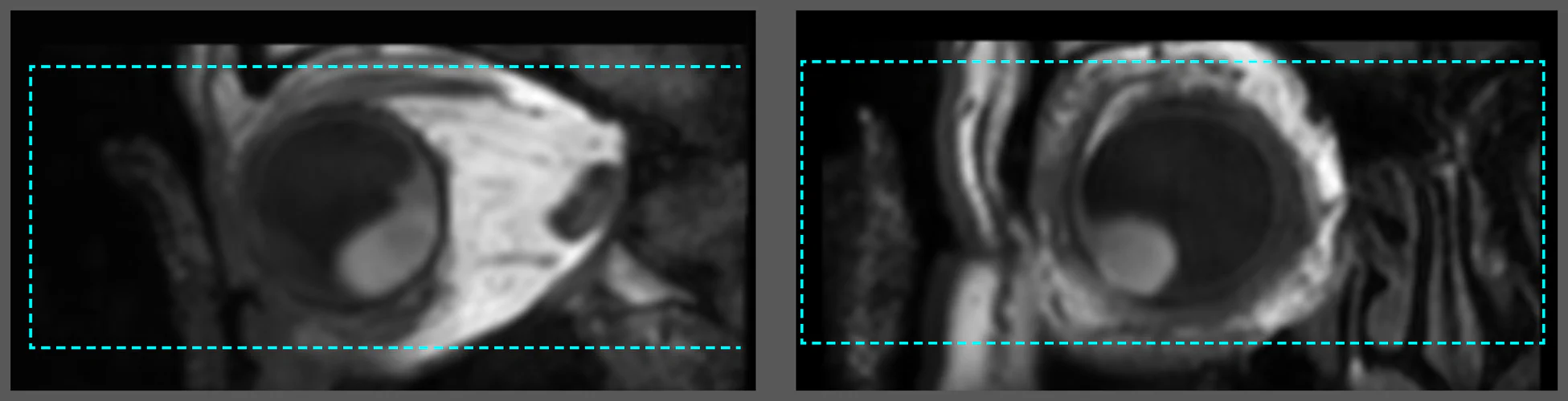
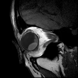
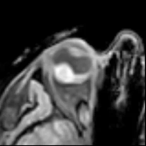
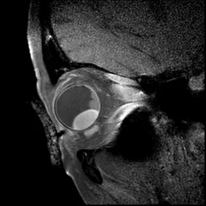
2D T2
The high in-plane resolution allows for accurate assessment of the lesions origin and relation.
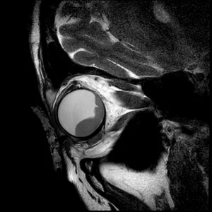
T1 & T1Gd
Contrast enhanced scans to differentiate the lesion and retinal detachment.
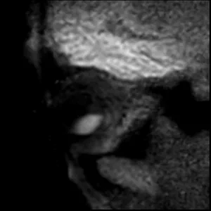
DWI ADC
Diffusion weighted imaging provides a valuable biomarker for differential diagnosis.
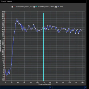
PWI
Perfusion weighted imaging provides a valuable information for differential diagnosis and of treatment response.
‹
›
Scans
| Sequence | Description | Geo | Time |
|---|
| Survey | Short survey covering both eyes | | 1:01 |
| 3D T2 | 3D T2-weighted scan with isotropic resolution | 3Dtra | 2:57 |
| 3D T1 | 3D T1-weighted scan with isotropic resolution | 3Dtra | 2:07 |
| MS T1 | T1-weighted multi-slice scan with high in-plane resolution | Obl | 1:16 |
| MS T2 | T1-weighted multi-slice scan with high in-plane resolution | Obl | 1:30 |
| DWI | Diffusion weighted scan with two b-values | Obl | 1:41 |
| 3D T1 SPIR | 3D T1 scan with isotropic resolution and fat suppression | 3Dtra | 2:07 |
| T1 mapping | Flip angle series and B1-mapping | Dyn | 0:21 |
| PWI | Dynamic scan with a 2 seconds temporal resolution | Dyn | 3:59 |
| 3D T1 SPIR Gd | 3D contrast-enhanced T1 scan with isotropic resolution | 3Dtra | 2:07 |
| MS T1 SPIR Gd | Contrast-enhanced T1 scan with high in-plane resolution | Obl | 1:22 |
Planning
3Dtra
Main geometry for the 3D sequences. As these scans have an isotropic resolution and can thus be reformatted in any orientation, they are scanned without angulation.
We plan them in the axial plane, with frequency encoding in the LR direction, to prevent fold-over artefacts.
Dyn
Same geometry as 3Dtra, but with slightly smaller FOV in the FH direction, to facilitate a higher temporal resolution for the dynamic scan.
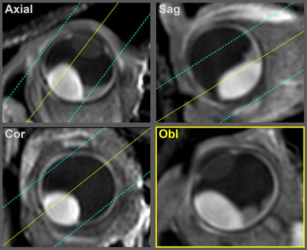
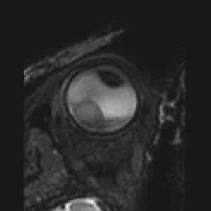
Obl
The orientation of multi-slice scans is tailored to the location and geometry of the tumour. It is typically perpendicular to the sclera and chosen such that the lesion and associated pathology are visualized in one slice.
For patients with complex pathology, e.g. both retinal detachment and optic nerve invasion, we perform these scans twice with different orientations.
Sequence details
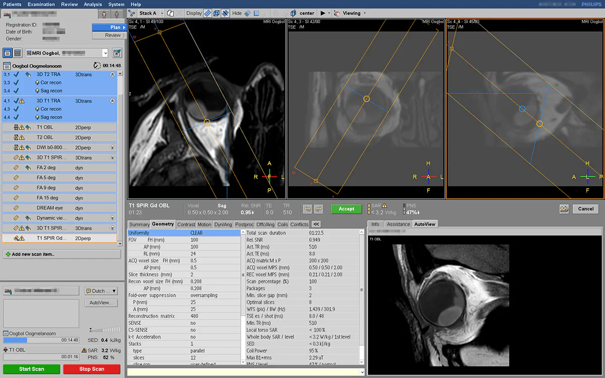
Download the Philips examcard (release 5.6.1.2), or view the complete list of scan parameters.
3D T2
An isotropic 3D T2-weighted sequence with fat supression, allowing for a 3D visualisation of the eye and surrounding anatomy.
Main sequence parameters:
Scan duration
02:57 min
Sequence type
3D TSE
TR/TE
2500 / 330 ms
ACQ voxelsize
0.78 x 0.80 x 0.80 mm
REC voxelsize
0.26 x 0.26 x 0.40 mm
WFS (pix) / BW (Hz)
0.74 / 588
TSE factor / shot length
117 / 616 ms
Fat saturation
SPIR
View the full sequence details.
3D T1
An isotropic 3D T1-weighted sequence, allowing for a 3D visualisation of the eye and surrounding anatomy.
Main sequence parameters:
Scan duration
02:07 min
Sequence type
3D TSE
TR/TE
400 / 31 ms
ACQ voxelsize
0.80 x 0.80 x 0.80 mm
REC voxelsize
0.36 x 0.36 x 0.40 mm
WFS (pix) / BW (Hz)
1.78 / 244
TSE factor / shot length
20 / 188 ms
View the full sequence details.
MS T1
A T1-weighted sequence with an increased in plane resolution, allowing for a better assessment of the origin of the lesion and its relation to the surrounding anatomy.
Main sequence parameters:
Scan duration
01:16 min
Sequence type
Multi-slice TSE
TR/TE
718 / 8.0 ms
ACQ voxelsize
0.50 x 0.50 x 2.00 mm
REC voxelsize
0.21 x 0.21 x 2.00 mm
WFS (pix) / BW (Hz)
1.60 / 272
TSE factor / shot length
6 / 48 ms
View the full sequence details.
MS T2
A T2-weighted sequence with an increased in plane resolution, allowing for a better assessment of the origin of the lesion and its relation to the surrounding anatomy.
Main sequence parameters:
Scan duration
01:30 min
Sequence type
Multi-slice TSE
TR/TE
1331 / 90 ms
ACQ voxelsize
0.40 x 0.42 x 2.00 mm
REC voxelsize
0.21 x 0.21 x 2.00 mm
WFS (pix) / BW (Hz)
2.25 / 193
TSE factor / shot length
17 / 180 ms
View the full sequence details.
DWI
A diffusion weighted sequence with two B-values (0 and 800 s/mm2) and ADC map. This sequence is particularly useful for differentiating between benign and malignant lesions.
Main sequence parameters:
Scan duration
01:41 min
Sequence type
Multi-slice TSE
TR/TE
1689 / 51 ms
ACQ voxelsize
1.25 x 1.42 x 2.40 mm
REC voxelsize
0.63 x 0.63 x 2.40 mm
WFS (pix) / BW (Hz)
0.688 / 631.3
TSE factor / shot length
Single shot / 492 ms
Fat saturation
SPIR
View the full sequence details.
3D T1 SPIR
An isotropic 3D T1-weighted sequence with fat supression. This sequence is particularly usefull when there is suspiccion of extra-scleral tumour extensions. In these patients it provides a reference to determine if the extra-ocular component is enhancing.
Main sequence parameters:
Scan duration
02:07 min
Sequence type
3D TSE
TR/TE
400 / 31 ms
ACQ voxelsize
0.80 x 0.80 x 0.80 mm
REC voxelsize
0.36 x 0.36 x 0.40 mm
WFS (pix) / BW (Hz)
1.78 / 244
TSE factor / shot length
20 / 188 ms
Fat saturation
SPIR
View the full sequence details.
T1 mapping
Flip angle series
Flip angle mapping sequence for T1 mapping. We use for flip angles (2, 5, 9 and 15 degrees) to accomodate the wide range of T1 values of intra-ocular masses.
Main sequence parameters:
Scan duration
00:07 min
Sequence type
3D GE
TR/TE
7.0 / 3.1 ms
Flip ange
2, 5, 9, 15 degrees
ACQ voxelsize
1.25 x 1.51 x 1.52 mm
REC voxelsize
1.00 x 1.00 x 1.00 mm
WFS (pix) / BW (Hz)
0.41 / 1059
Fat saturation
PROSET 11
View the full sequence details.
B1-mapping
B1 mapping using the DREAM sequence. This sequence is used to determine the actual flip angle for the T1 mapping and PWI analyses.
Main sequence parameters:
Scan duration
00:14 min
Sequence type
MS GE
TR/TE
7.1 / 2.3 / 3.6 ms
ACQ voxelsize
2.00 x 2.00 x 2.00 mm
REC voxelsize
1.00 x 1.00 x 2.00 mm
TFE dur. shot / acq (ms)
466.5 / 428.2
TFE shot interval (ms)
6997
WFS (pix) / BW (Hz)
0.40 / 1085
View the full sequence details.
PWI
Dynamic Contrast Enhanced sequence for perfusion analysis of intra-ocular masses. TWIST is used to increase the temporal resolution.
Main sequence parameters:
Scan duration
03:59 min
Sequence type
3D GE
TR/TE
4.5 / 2.3 ms
Flip angle
13 degrees
ACQ voxelsize
1.25 x 1.51 x 1.52 mm
REC voxelsize
1.00 x 1.00 x 1.00 mm
WFS (pix) / BW (Hz)
0.39 / 1116
Fat saturation
PROSET 11
Dyn. scan time
00:01.9 s
Central/Periph. size
25% / 20%
View the full sequence details.
3D T1 SPIR Gd
3D T1-weighted sequence with fat supression after gadolinium contrast administration. This sequence is used to determine the enhancement pattern of the intra-ocular masses. The sequence parameters are the same as the 3D T1 SPIR sequence.
Main sequence parameters:
Scan duration
02:07 min
Sequence type
3D TSE
TR/TE
400 / 31 ms
ACQ voxelsize
0.80 x 0.80 x 0.80 mm
REC voxelsize
0.36 x 0.36 x 0.40 mm
WFS (pix) / BW (Hz)
1.78 / 244
TSE factor / shot length
20 / 188 ms
Fat saturation
SPIR
View the full sequence details.
MS T1 SPIR Gd
T1-weighted sequence with fat supression after contrast, the increased in plane resolution allows for a better assessment of the lesions relation to the surrounding anatomy.
Main sequence parameters:
Scan duration
01:22 min
Sequence type
Multi-slice TSE
TR/TE
510 / 8.0 ms
ACQ voxelsize
0.50 x 0.50 x 2.00 mm
REC voxelsize
0.21 x 0.21 x 2.00 mm
WFS (pix) / BW (Hz)
1.60 / 271
TSE factor / shot length
6 / 48 ms
Fat saturation
SPIR
View the full sequence details.
 3DT1 SPIR GdThe isotropic resolution allows for accurate 3D assessment of the lesion dimensions.
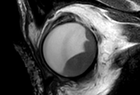 2D T2The high in-plane resolution allows for accurate assessment of the lesions origin and relation.
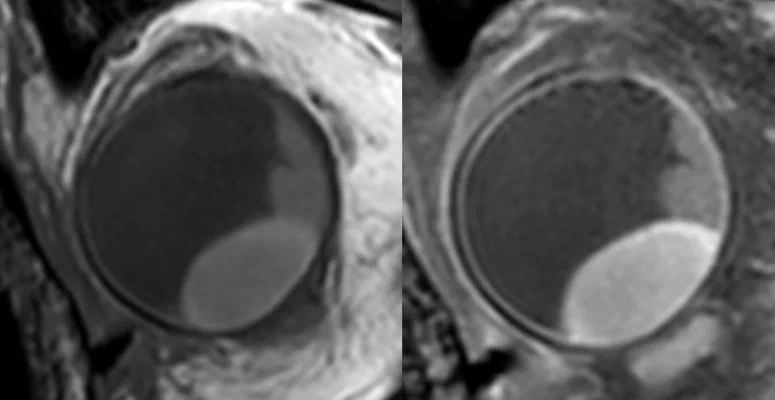 T1 & T1GdContrast enhanced scans to differentiate the lesion and retinal detachment.
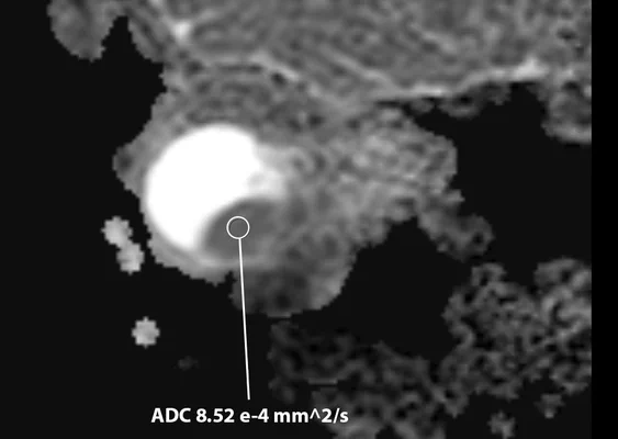 DWI ADCDiffusion weighted imaging provides a valuable biomarker for differential diagnosis.
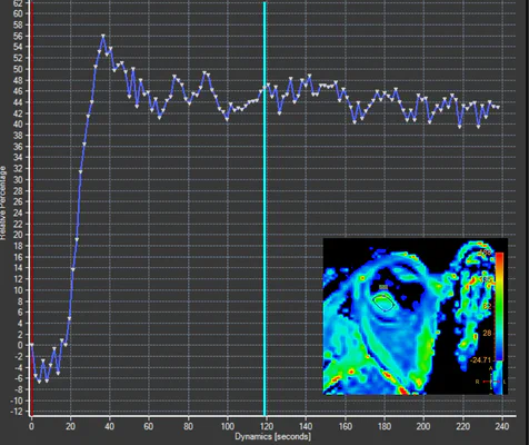 PWIPerfusion weighted imaging provides a valuable information for differential diagnosis and of treatment response.